The human body is a complicated structure. Proper diagnosis and treatment may depend on knowing what’s happening underneath the surface. It is where the amazing range of technology known as medical imaging comes in, enabling doctors to look right into your body. Say goodbye to the days of depending just on old black-and-white X-rays! With the help of this thorough book, you’ll be able to navigate the field of medical Imaging Solutions.
We’ll examine the most effective healthcare modalities and discover how each technology functions. Learn about the physics underlying these wonders, from using magnetic fields to create precise 3D pictures to using sound waves to capture peeks in real time. Find detailed information about the advantages and disadvantages of each imaging test. Also, find the necessity of making well-informed inquiries during healthcare consultations. This guide is your one-stop shop for learning about the marvels of diagnostic imaging, whether you’re a curious patient looking for clarification or just someone captivated by medical developments!
Table of Contents
ToggleHealthcare Imaging Modalities and their Applications
Ultrasound

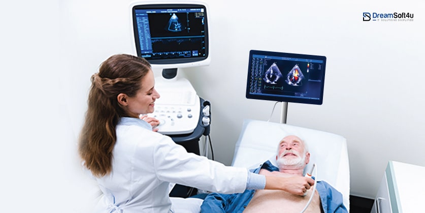
Ultrasound is becoming a necessity in the healthcare industry as it provides a deep examination of the human body and diagnoses internal issues. Almost every integrated healthcare system relies on ultrasound or sonography machines to examine the patient and provide suitable medication. Ultrasound uses high-frequency sound waves instead of ionizing radiation like X-rays do to provide real-time pictures of soft tissues and organs.
Functionalities:
The fundamental ultrasonic imaging solutions technique is comparable to submarine sonar. The ultrasonic machine sends high-frequency sound waves that are inaudible to human ears. The equipment records the echoes that return after these waves pass through the body and are reflected by interior structures. The ultrasound instrument makes real-time viewing of organs and tissues possible by analyzing these echoes and converting them into intricate visual representations on a screen.
Its Uses:
Ultrasound is an invaluable tool for a variety of diagnostic procedures due to its versatility. It can penetrate well through the abdominal wall thereby giving a clear view of vital organs like the liver, kidneys etc. When it comes to pregnancy care (obstetrics) and female reproductive health (gynaecology), this method cannot be overlooked as far as monitoring fetal growth alongside assessing the uterus and ovaries’ condition is concerned among others. Additionally, hospital management systems diagnose problems related to muscles or bones; guiding biopsy procedures towards lumps that may be cancerous and thus should not be left out while dealing with different types of diseases but how does it work? Well, basically by shows us how blood circulates within our blood vessels using special kinds of waves.
Benefits:
The benefits of ultrasonography are numerous and appealing. Clinicians may observe the movements of organs like a beating heart or developing baby since ultrasonography records in real-time. This dynamic portrayal offers crucial insights into organ function that are not feasible to gain from static pictures.
Limitations:
Although it’s a fantastic tool, ultrasound has a few disadvantages. What can be observed is limited because sound waves cannot easily travel through air-filled tissues like the lungs or bone. The quality of the picture may also be impacted by the operator’s skill and knowledge of the ultrasonic probe. Adipose tissue or overlapping organs may occasionally hide deeper structures, requiring other Imaging Solutions modalities to obtain a more thorough assessment.
CT Scan

CT scan is a workhorse for medical imaging. It produces finely detailed cross-sectional pictures of your body using sophisticated computer processing and X-ray technology. Like slicing a loaf of bread, a CT scan creates several thin “slices” of your insides that medical professionals may stack to create a complete three-dimensional image.
Functionalities:
The CT scan technology provides fine details about blood arteries, soft tissues, and bones. It takes many pictures of your body from various angles by rotating an X-ray source around it. Subsequently, powerful computers create a very detailed three-dimensional representation of these photographs.
Its Uses:
CT scans are invaluable in a wide range of situations. With the help of this machine, Doctor can detect the internal injuries and tumours of an individual evaluating blood flow and bone detail. It is crucial in diagnosing cancer, heart disease, and musculoskeletal conditions.
Most Preferred Software Development Company Since 2003
Request A Free Quote
98% Client Retention
Benefits:
CT Scan produces high-resolution pictures that enable more precise diagnosis in comparison to X-rays. They’re also quick and painless, so multiple patients can benefit from them.
Limitations:
While powerful, CT scans do have limitations. It exposes patients to ionizing radiation at a higher dose than X-rays. In some cases, contrast dye injections may be required, which can cause side effects. Additionally, CT scans may not suit everyone due to factors like claustrophobia or certain medical implants.
Fluoroscopy

Fluoroscopy is an automated Imaging Solutions producing machine using a low beam of X-ray to generate the time structure and physiology of an individual. Although it may look similar to the X-ray machine, it targets the deep tissues and identifies the anatomical problem in the body. People can identify fluoroscopy as a traditional way of X-ray, capturing tools that dynamically visualize organs and tissues along with their motion to develop a fluoroscopic image.
Functionalities:
Fluoroscopy machines Bombard the X-ray projection on the particular section of the body where doctors need to diagnose the patient. The X-ray beam enlightens the particular section of the body and the special detector in the machine captures the image of the cross-section. With the help of this machine, Doctors can also detect the organ tissue and bones of an individual in the form of images and videos.
It’s Uses:
Fluoroscopy is helpful in circumstances when mobility is crucial. It allows physicians to observe how food passes through your intestines and throat, sort of like a detective show for your digestive system. It can also examine the movement of your heart, lungs, and joints in a normal state. Fluoroscopy provides a real-time image of the inside of the body to assist doctors in guiding specialized treatments such as biopsies or catheter threading.
Benefits:
Medical professionals may see things more accurately in real-time with the help of fluoroscopy that they could miss with X-ray. It can result in more precise operations and even more accurate diagnosis. Consider it a very useful and educational addition to your physician’s toolkit. Nowadays mhealth apps are also utilized to store the necessary data by the hospitals and patients.
Limitations:
Fluoroscopy involves some X-ray exposure. Therefore doctors do not recommend this examination frequently or in routine checkups. Additionally, it also requires dye which may have side effects on body parts or an individual. Since it involves radiation, it’s best used when other imaging techniques won’t cut it. Fluoroscopy can be a real game-changer for understanding what’s happening inside you.
Radiography

Radiography is also popularly known as X-ray imaging. It utilizes a burst of ionizing radiation to capture a still image of your internal structures. It’s just like taking a picture of a particular section of your bone or denser tissue. It is a quick and painless procedure that allows doctors to see fractures, and joint abnormalities, and even detect pneumonia or foreign objects within the body.
Functionalities:
A regulated X-ray beam is directed toward the desired body part using this technique. X-ray penetrates different sections of the body at different speeds and wavelengths so that it can penetrate the diagnostic section of the part and provide accurate results. Eventually, the majority of X-ray beams are directly observed by the harder tissue of bones and can’t penetrate the deep tissue of bones. The remaining X-rays are collected by a specialized detector behind the subject, and they are then processed into a black-and-white picture.
Its Uses:
Radiography is an essential diagnostic tool for a wide range of medical disorders. It provides a flexible diagnostic tool that may be used to expose pneumonia in the lungs, discover foreign items within the body, uncover shattered bones, and detect joint abnormalities. It serves as a cornerstone for first assessments and frequently serves as the basis for additional research.
Benefits:
There are some advantages to radiography. It’s a quick and painless process that works for a variety of folks. In contrast to other imaging solutions, it is usually more accessible and less expensive in medical settings. Moreover, X-ray Imaging Solutions produces an enduring record that is readily shared and saved for later use.
Limitations:
Radiography has limits even if it is useful. It is best for rendering thick tissues and bones. Soft tissues, such as muscles and organs, frequently show less clarity and may need further imaging to provide a conclusive diagnosis. Ionizing radiation is given to patients during X-rays, albeit the dosage is often minimal for routine radiography operations.
MRI

Magnetic Resonance Imaging (MRI) is a non-invasive imaging of the body. This machine is used to examine the deep tissue and organs of the body to find the problem. MRI works on radioactive waves which have strong magnetic fields that produce an impression of a particular section of the body so that Doctors can examine the illness. Generally, medics use this modality to diagnose tumours or severe injuries in the internal organs of the body.
Functionalities:
While examining through the MRI machine the patient is surrounded by a strong magnet that produces a strong magnetic field to align the protons and tiny particles of the body. After that, certain frequencies of radio waves are pulsed, which causes these protons to absorb energy and realign. The MRI scanner picks up the energy they emit when they realign to their initial form. High-resolution cross-sectional pictures may be produced because of the intricate interaction between radio waves and magnetism.
Its Uses:
MRI is commonly used to diagnose the internal body parts and growth in the tissues. If a patient has any unnecessary growth inside the body or has any damaged organs, then an MRI machine can easily detect the issue, and the Doctor can coordinate with the patient accordingly.
Benefits:
MRI makes it possible to identify anomalies and make diagnoses with greater accuracy. Because routine MRI scans are painless and non-invasive, they may be performed on a wider variety of patients than treatments that need needles or contrast dye injections, however, these techniques are occasionally employed in conjunction with MRIs. MRI is a highly flexible diagnostic technique that can evaluate almost any area of the body.
Limitations:
MRI scans use high magnetic fields and energy, so it could be expensive for some of the users. MRI produces more effective results than a CT scan therefore, it has a massive impact on the body that can damage the body if exposed for long. For certain people, the confined aspect of the MRI scanner may cause claustrophobia. Owing to safety issues, some metallic implants or medical equipment could not work with magnetic resonance imaging (MRI).
Echocardiography

A quick heart rate diagnostic gauge is termed an echocardiography machine. This tool detects the electrical signal generated by the pumping of the heart and develops a Graphical image representing the pressure and pumping of the heart. Echocardiography works on the principle of pulsating sound produced by the pumping of blood into nerves. Overall, this tool is used to diagnose the status of the heart and its condition in different situations or illnesses.
Functionalities:
A heartbeat produces tiny electrical currents that are detected by tiny electrodes affixed to the skin on selected parts of the body. Now the ECG machine receives these electrical signals and amplifies them to draw the frequency of heartbeat to diagnose the status of the patient.
Its Uses:
ECGs are generally used to diagnose the different types of cardiac disorders. Doctor generally uses this equipment to diagnose the heart rate of the patient. In some cases, they are also used during the operation so that they can keep the status of the heart rate and prevent any failure or collapsing situations.
Benefits:
Since there are no needles or injections required, a variety of people can benefit from them. Generally speaking, the process is inexpensive and rapid in comparison to other cardiac diagnostic procedures. ECGs provide important information on cardiac rhythm and function.
Limitations:
ECGs only record a split second and might not always pick up on sporadic anomalies. To spot such problems, an experienced medical practitioner has to evaluate the ECG readings. For a conclusive diagnosis, ECGs may need to be combined with other investigations.
Angiography

Angiography is detailed imaging of the circulatory system and the veins of the body to detect any blockage or irregularities in the patient. It is commonly used in the diagnosis of blocked veins or networking that might affect the paralytic attack or tubal blockage. During angiography, doctors make small incisions and with the help of surgical microscopes, they can fix the problem.
Functionalities:
Angiography utilizes X-ray Imaging Solutions with a unique twist. A thin, catheter-like tube is inserted into a blood vessel. This tiny tube acts as a delivery system for a contrast dye. The dye travels through your bloodstream, illuminating your arteries and veins on the X-ray images. By capturing images from various angles, doctors can build a comprehensive picture of your vascular network.
Its Uses:
This detailed map created by angiography is a game-changer for diagnosing vascular problems. Blockages, narrowings (atherosclerosis), aneurysms (bulges), and malformations in your blood vessels can all be identified with this technique. Think of a doctor needing to pinpoint the exact location of a blockage before opening it up – angiography provides that crucial roadmap. This information is vital for guiding treatment decisions in conditions like coronary artery disease (blocked arteries in the heart) and peripheral arterial disease (blocked arteries in the legs).
Benefits:
Angiography offers several advantages over traditional methods as it provides provision and a clear picture of the blood vessels which makes it easy to diagnose. Additionally, the decision made during the angiography can be recovered within no time.
Limitations:
Although it is the best procedure to diagnose the internal problems in the veins and networking system, it has some limitations that cannot be diagnosed with angiography. While operating if the doctor does not make a precise cut, then it can lead to complete rupture of the veins and blood vessels. Additionally, an internal organ of the body is in direct exposure to harmful radiation.
Get your fully optimized Software for Different Modalities
Reach out the top professionals of DreamSoft4u
Necessity of Imaging Modalities
Human bodies are complex structures and their anatomical issues cannot be identified by a simple physical examination or patient history. Nowadays the modern technique also involves Mobile healthcare apps to keep track of patient’s data. During diagnosis medical professionals use imaging techniques to see hidden structures and anomalies in the body. Imaging solutions provide a blueprint of the human body that is comparable to what a technician would require to identify flaws in your car’s engine.
MRIs, CT scans, X-rays, and ultrasounds are just a handful of the many imaging modalities that are accessible. Every modality is particularly good at rendering particular tissues and structures, offering insightful information about possible problems. Imaging solutions provide a thorough diagnostic approach, encompassing everything from identifying cancers and fractures to analyzing internal bleeding and assessing organ function. In the end, they enable doctors to identify patients accurately, which results in better treatment regimens and patient outcomes.
Role of Software Development Company in Imaging Techniques
Can you imagine having to service an engine on a car without being able to look inside? That was the situation confronted by medical experts prior to the development of advanced imaging technology. Software development companies are the secret weapon behind these fantastic tools.
Think of a fancy CT scanner – it captures a ton of data, but that data’s useless without special software to turn it into clear pictures. The tools that take that raw data and turn it into intricate visuals that physicians can use to detect issues are created by these development teams. These are the wizards working behind the scenes to transform those hazy blobs into incredibly clear images of your inside organs.
But software developers continue to make pretty pictures. They also create programs to manage and analyze all those images. Imagine a giant library of medical scans – these companies build systems that let doctors securely store, find, and share patient images easily. Furthermore, advanced analytical techniques utilizing artificial intelligence to assist physicians in identifying issues more quickly and precisely. Therefore, keep in mind the software engineers who are working behind the scenes to assist doctors in understanding what’s occurring inside of you and provide you with the therapy you need the next time you receive an X-ray or other scan.
Wrapping Up!
Technology developments promise increasingly more advanced tools for patient diagnosis and treatment, which bodes well for the future of medical imaging. Leading the charge in this advancement is healthcare software development. Software developers are making imaging solutions modalities completely usable for healthcare practitioners by offering user-friendly interfaces, strong image processing algorithms, and secure data management systems. It leads to improved treatment strategies, quicker and more accurate diagnoses, and ultimately better patient outcomes. Custom healthcare software is an investment in the medical industry’s future. It ensures that sophisticated imaging technologies will be effectively used to improve the quality of life for a great number of patients.
FAQs
Q1 What imaging test is right for me? (X-ray vs. CT vs. MRI)
The best Imaging Solutions depend on your specific situation. X-rays are quick and good for bones, while CT scans offer more detail for internal injuries. MRIs excel at examining soft tissues like muscles and the brain. Your doctor will recommend the most suitable option.
Q2 Is radiation exposure a concern?
Radiation is used in several imaging procedures (such as CT scans and X-rays). To reduce exposure, clinicians adhere to stringent guidelines (ALARA). In certain cases, individuals may experience allergic responses when contrast dye is employed for improved visibility. Address any worries in advance with your physician.
Q3 What’s new in the world of imaging technology?
AI in medical imaging is gaining popularity! With the use of AI algorithms, physicians can diagnose patients more quickly and correctly by analyzing photos. Furthermore, CT scans are developing to produce pictures of greater quality while requiring fewer radiation doses. Advanced tools for better patient care will be the main focus of imaging solutions in the future.


